Revolutionizing
Human Disease Research
and Diagnostics

Groundbreaking EV isolation technology enabling minimally invasive human disease screening, diagnosis, and treatment monitoring.

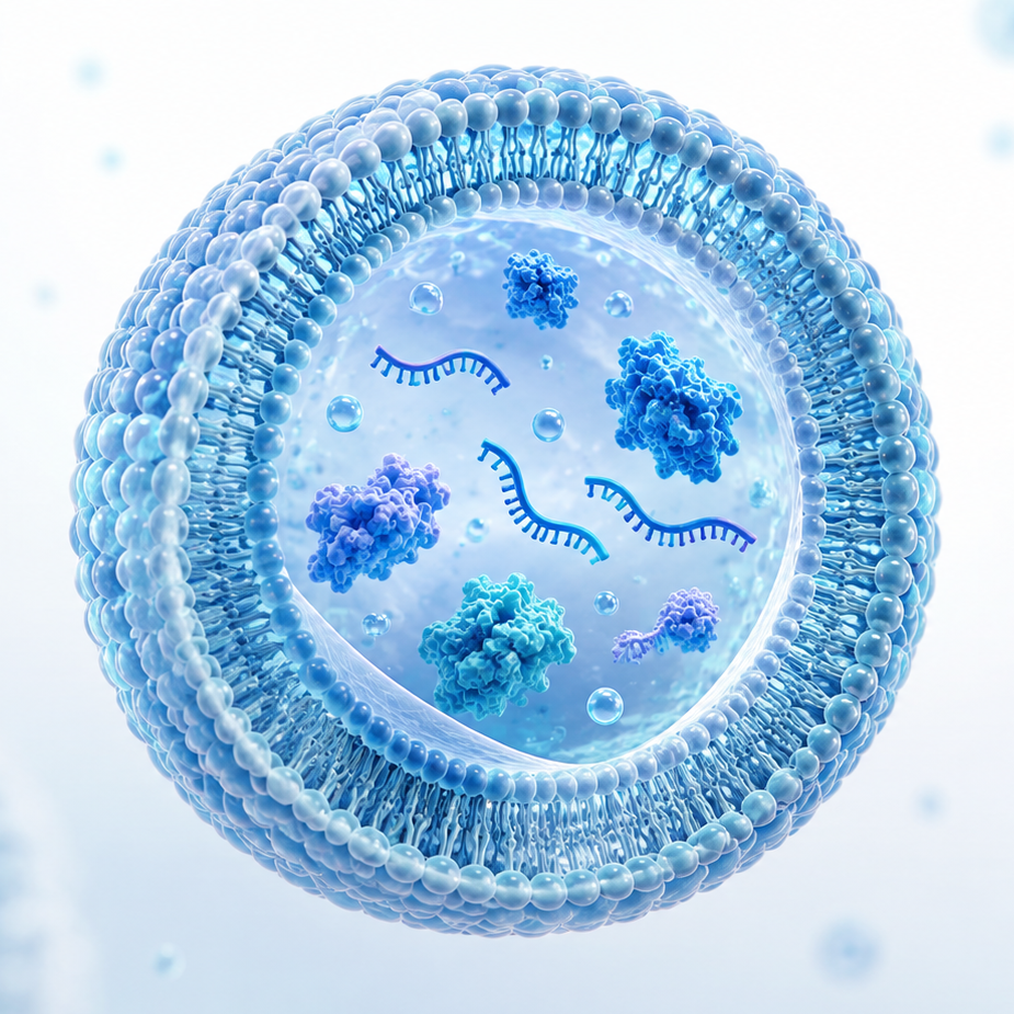
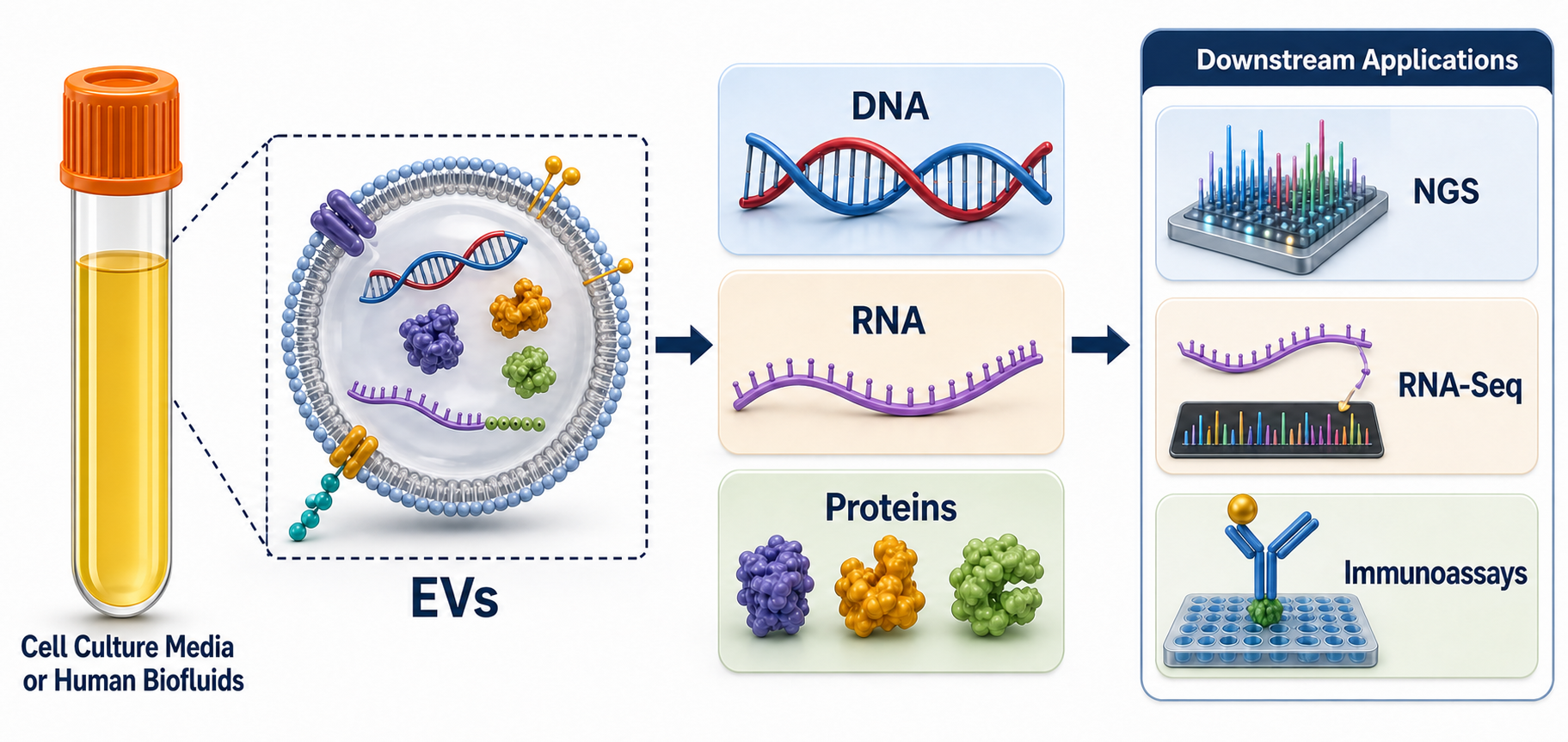
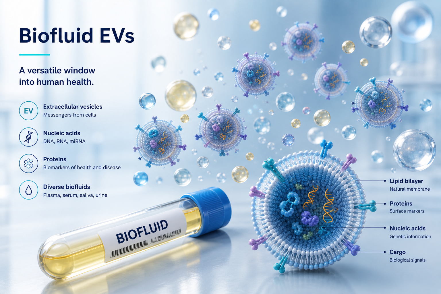
Extracellular vesicle carrying DNA, RNA, and protein cargo

Enabling rapid liquid biopsy

For decades, understanding disease at the molecular level has meant invasive tissue biopsies — painful to obtain, hard to repeat, and often impossible to access safely. Captis is rewriting that standard. Every cell continuously releases extracellular vesicles into blood and other bodily fluids, carrying the same tumor-derived DNA, RNA, and protein cargo found in solid tissue. By capturing and analyzing these vesicles, we turn a simple blood draw into a window on disease — enabling diagnostics that are minimally invasive, repeatable, and fast enough to keep pace with how a patient is actually doing.

Mission & Vision

Mission
Advancing diagnostics through nanotechnology

Our primary mission at Captis Diagnostics is to spearhead the advancement and commercialization of cutting-edge research tools for human disease research and clinical diagnostics, anchored by our Lipid Nanoprobe (LNP) platform, which enables rapid, high-efficiency extracellular vesicle isolation from human biofluids.

LNP magnetic isolation
>90%
LNP EV capture efficiency in plasma
15 min
Rapid, simple LNP isolation workflow
Vision
Empowering researchers and clinicians alike

Captis Diagnostics envisions a future where our EVs-based liquid biopsy technology empowers researchers and healthcare providers to create minimally invasive diagnostic tools and assays for human diseases. These innovative diagnostic assays will provide medical doctors with actionable insights for treating human diseases, ultimately leading to improved patient outcomes.

Vision

Our Lipid Nanoprobe (LNP) Technology

Lipid nanoprobe (LNP) EV isolation technology

Isolation principles
I. Isolation principles

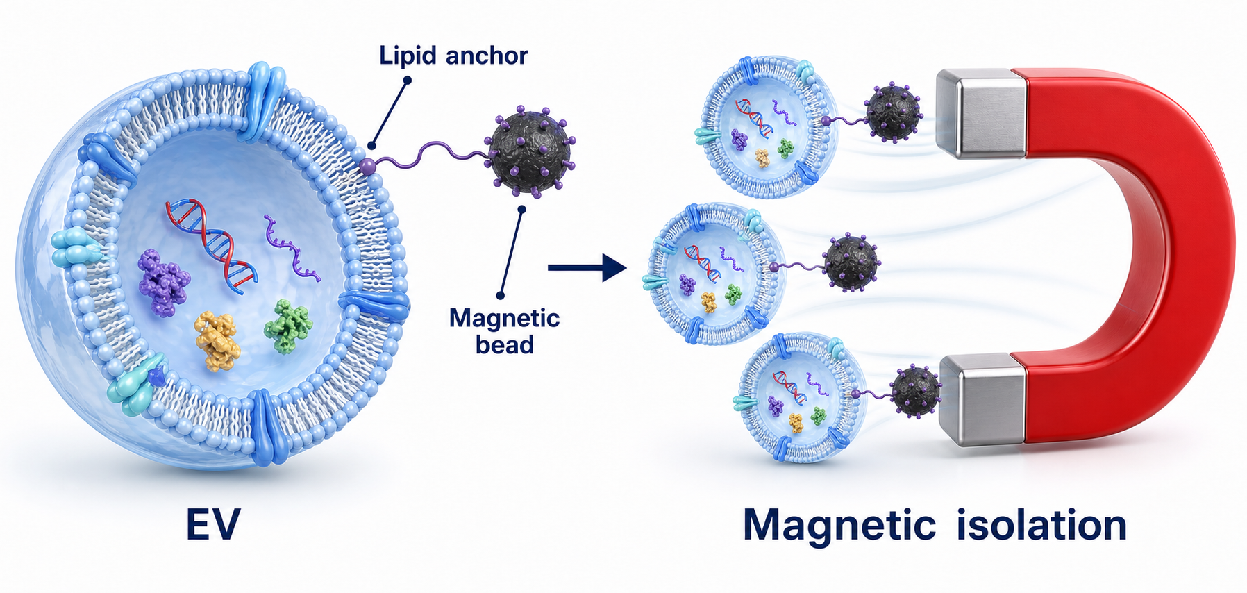
Targeting the lipid bilayer

LNP leverages the fundamental structural characteristic of EVs — their outer bilayer lipid membrane — to isolate them with unmatched efficiency and specificity.

The LNP system uses two nanomaterial components: a lipid-based labeling probe (lipid-PEGm-biotin) that inserts into the lipid membrane through self-assembly to instantly tag EVs, and magnetic nanospheres (MNS) surface-modified with NeutrAvidin to collect the tagged EVs magnetically.

Read the original LNP paper →
LNP isolation workflow
II. Sample-to-isolation workflow

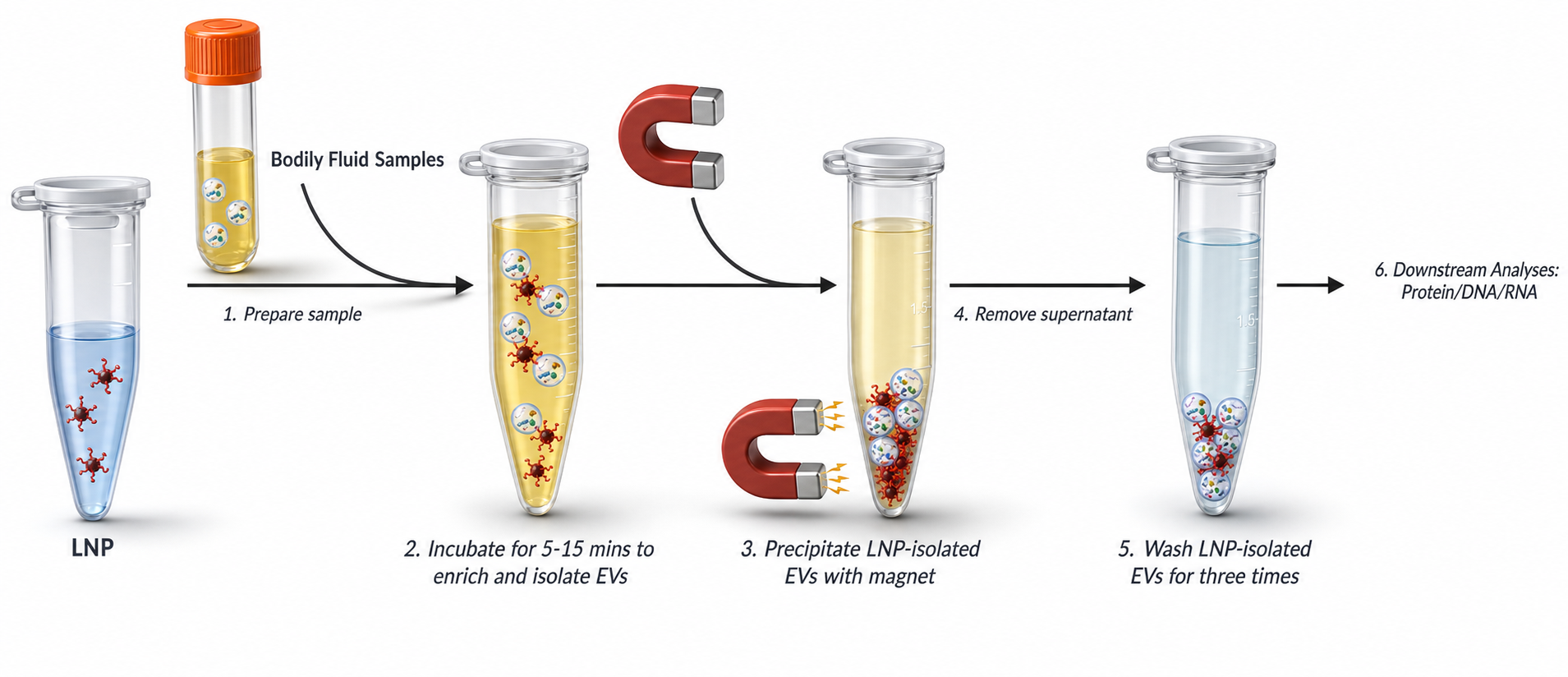
A simple six-step protocol

Starting from a raw bodily fluid sample, LNP isolates EVs through a single magnetic separation — no ultracentrifugation or specialized equipment required.

After a short incubation, LNP-bound EVs are magnetically precipitated, washed, and prepared for downstream protein, DNA, and RNA analysis — the full workflow takes 15 minutes or less.

LNP omics workflow
III. LNP-enabled EV omics workflow

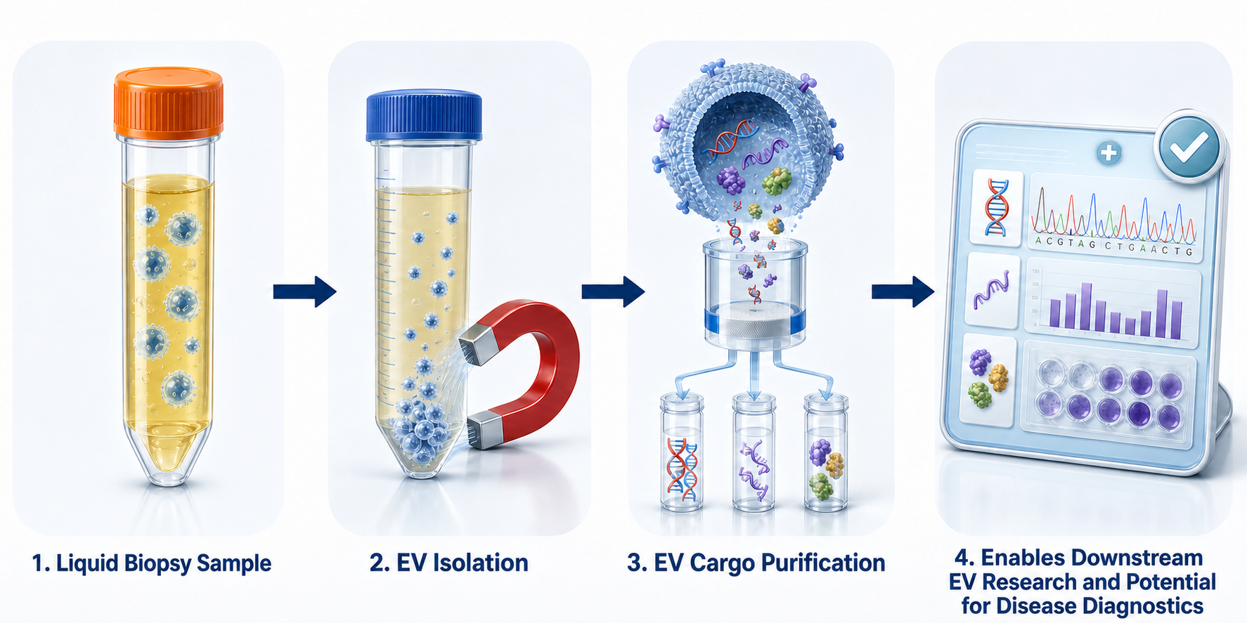
From sample to multi-omics insight

LNP-isolated EVs harbor high-quality nucleic acids (DNA and RNA) and unique proteins, supporting DNA mutation detection, RNA sequencing, and proteomics from a single blood draw.

Reference: Y. Wan, et al. Rapid magnetic isolation of extracellular vesicles via lipid-based nanoprobes. Nature Biomedical Engineering 1 (2017) 0058.

What sets LNP apart

Total EV isolation efficiency
Over 90% EV capture from plasma — unmatched across competing methods including ultracentrifugation and precipitation.
Rapid, simple workflow
Rapid, simple protocol completed in less than 15 minutes. No bulk machine required. Compatible with automated liquid handling systems.
Downstream multi-omics
LNP-captured EVs harbor high-quality nucleic acids and unique proteins — compatible with DNA, RNA, and proteomics workflows.

Diagnostic Applications

Clinical utility in NSCLC

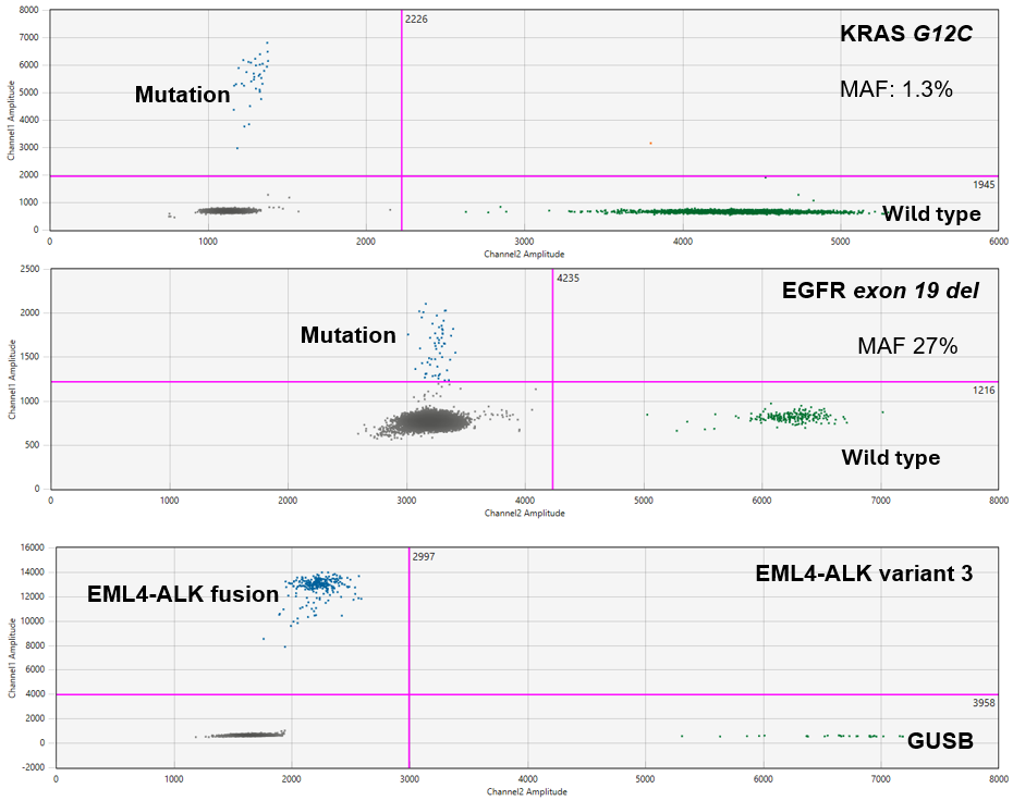
LNP-isolated extracellular vesicles carry actionable tumor-derived DNA directly from patient plasma, enabling non-invasive genomic profiling even when tissue biopsy is difficult to obtain or repeat. In a cohort of non-small cell lung cancer (NSCLC) patients, LNP technology was used to isolate EVs from a single blood draw and interrogate them for clinically relevant alterations.

DNA point mutations (KRAS, EGFR) and gene fusions (ALK-ELM4) were successfully detected in LNP-isolated EVs from plasma of non-small cell lung cancer patients, demonstrating clinical-grade sensitivity for actionable genomic alterations. Across the 13 patients tested, EV-based results matched tissue biopsy findings in all but one case, supporting plasma EVs as a reliable, minimally invasive alternative for genomic alteration detection.

Diagnostic application data
PatientMutationTissuePlasma EV
KRAS Mutation
1G12CMUMU
2G12CMUMU
3G12SMUMU
4G12SMUMU
5G13CMUMU
6G12AMUWT
7Q61HMUMU
EGFR Mutation
8EGFR L858RMUMU
9EGFR L858RMUMU
10EGFR Exon19 delMUMU
EML4-ALK fusion
11Variant 3a/bPositivePositive
12Variant 3a/bPositivePositive
13Variant 3a/bPositivePositive

KRAS dynamics in pancreatic cancer

In a prospective study of 44 patients with KRAS-mutated pancreatic ductal adenocarcinoma (PDAC) undergoing neoadjuvant therapy and surgery, LNP-isolated plasma EVs were used to track KRAS mutation allele frequency (MAF) over the course of treatment. Patients with rising EV-KRAS MAF had significantly shorter disease-free and overall survival than those with stable or decreasing MAF, and this trend independently predicted survival outcomes on multivariable analysis.

Read the full paper →

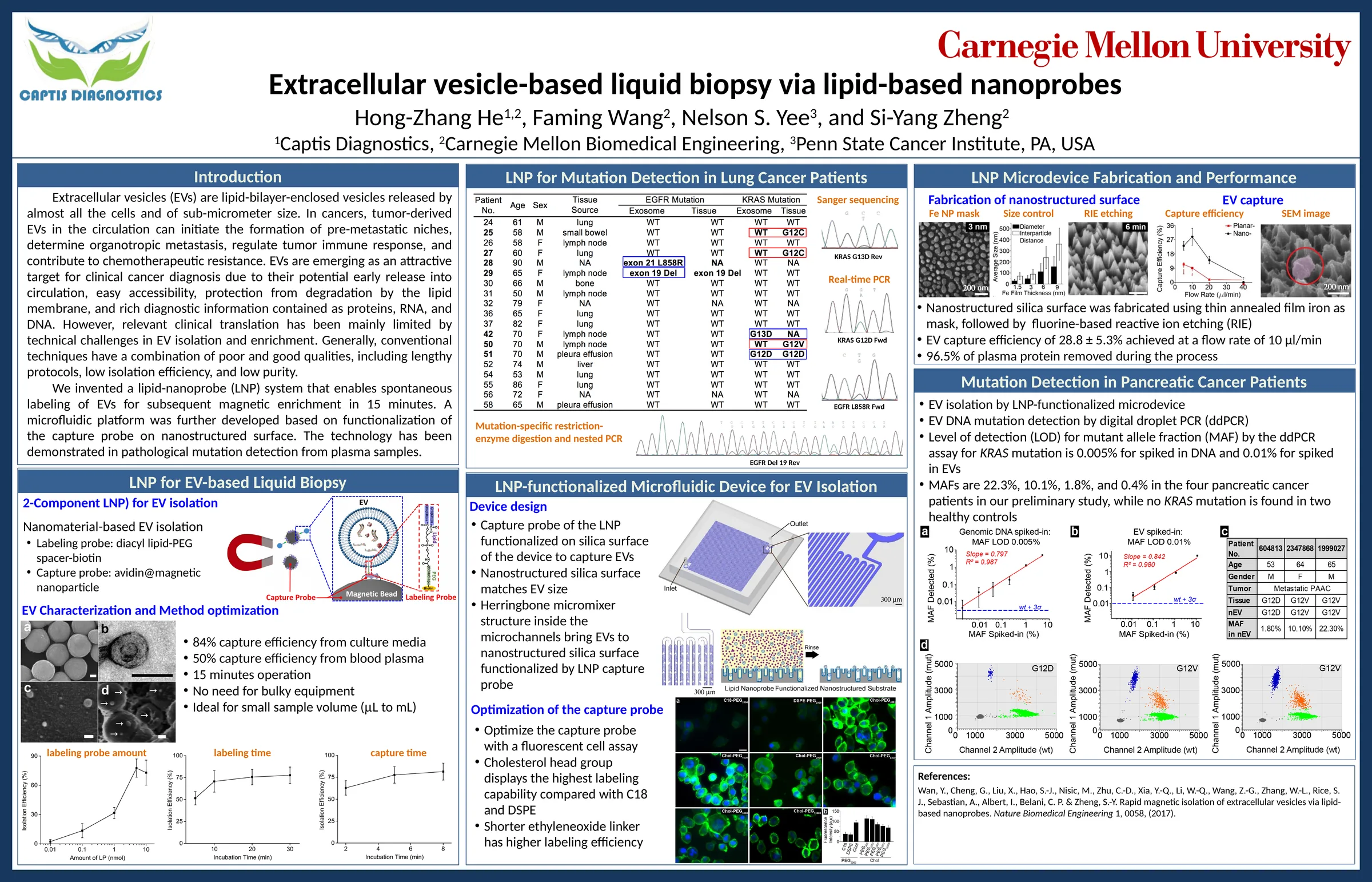
Extracellular vesicle-based liquid biopsy via lipid-based nanoprobes

Extracellular vesicle-based liquid biopsy via lipid-based nanoprobes conference poster

Relevant Publications

Precision EV Kits for
Every Workflow

Two purpose-built isolation kits — one optimized for multi-omics discovery, one for in-depth proteomics and mass spectrometry. Both powered by Captis's proprietary magnetic bead technology.

Protocol overview
Workflow
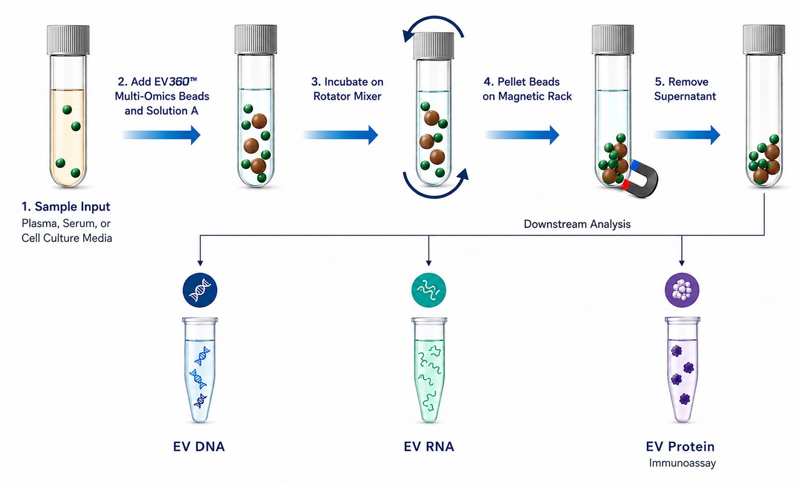
EVs360 Multi-Omics Kit protocol schematic
Product highlights
Rapid workflow
As little as 15 minutes for protein applications — no ultracentrifugation required
High recovery & yield
Over 90% EV capture efficiency from plasma using lipid nanoprobe technology
Versatile samples
Compatible with plasma, serum, and conditioned cell culture media
Quantitative & reproducible
Consistent inter- and intra-run performance for translational studies
Automation ready
High-throughput compatible with HulaMixer or liquid handling systems
Order options
10 reactions
EVs360™ Multi-Omics Kit
Cat. no. L10001 · 10 rxn
List price$550.00
Sample typesPlasma, serum, cell culture
For useResearch use only
Compatible downstream workflows
EV DNA isolation for genomic and epigenetic analysis
EV RNA isolation for transcriptomic profiling
EV protein isolation for immunoassay analysis
Frequently Asked Questions

Practical guidance for EV isolation and downstream DNA, RNA, and protein workflows.

What makes the EVs360™ Multi-Omics Kit different from ultracentrifugation?
The kit uses a magnetic bead-based workflow with a 15-minute EV capture step. It does not require an ultracentrifuge and is designed to support consistent processing across operators, low sample volumes, and multi-sample studies.
Does the kit isolate both exosomes and larger extracellular vesicles?
Yes. The workflow provides broad, pan-EV capture rather than selecting a single EV subtype. The recovered population may include small EVs commonly described as exosomes, microvesicles, and other membrane-bound EVs. EV composition can vary with sample type and pre-analytical handling.
Which sample types are supported?
The standard workflow is designed for plasma, serum, and cell culture media. Other biofluids — including urine, saliva, and cerebrospinal fluid — may be feasible, but bead amount, sample input, and wash conditions should be optimized and analytically validated before routine use.
Can frozen samples be used?
Yes. Frozen plasma, serum, and conditioned media can be used. Thaw samples completely, mix gently until homogeneous, and avoid repeated freeze-thaw cycles. If aggregates or debris are present, use the pre-clear option described in the Operator Manual.
Does the workflow require specialized equipment?
No ultracentrifuge is required. Standard laboratory equipment is sufficient:
  • Magnetic stand or rack
  • Tube rotator or mixer
  • Vortex mixer and calibrated pipettes
  • Standard microcentrifuge
  • Heating block or water bath when required by the downstream extraction method
Which multi-omics applications are compatible with the isolated EVs?
EV-bound material can support multiple molecular workflows after appropriate lysis and extraction, including:
  • DNA mutation, copy-number, methylation, and sequencing assays
  • RNA expression, gene-fusion, miRNA, qPCR/ddPCR, and sequencing workflows
  • Protein biomarker assays such as Western blotting, ELISA, Olink®, NuLISA™, and other immunoassays
Compatibility and analytical performance depend on the downstream kit, lysis chemistry, input requirements, and assay validation. Metabolomics and lipidomics workflows require application-specific optimization.
How should DNA and RNA be recovered from captured EVs?
Captured EVs may be lysed directly on the beads using a compatible commercial DNA or RNA extraction workflow. Use the EVs360™ supplementary DNA or RNA extraction protocol as the starting point, and follow the extraction-kit manufacturer's requirements for binding, washing, elution, and input volume.
Can the kit be used for protein analysis?
Yes. The Multi-Omics Kit can support Western blotting, ELISA, Olink®, NuLISA™, and other protein biomarker workflows after compatible lysis. For deep LC-MS/MS proteomics, use the dedicated EVs360™ Proteome-MS Kit (Cat. no. L20001 · 10 rxn or L20002 · 25 rxn), which is optimized for mass-spectrometry workflows.
Can one EV preparation be divided across DNA, RNA, and protein assays?
Potentially, but the experimental design must account for the input required by each assay. For low-abundance samples, splitting one preparation may reduce material below an assay's detection threshold. When possible, use matched captures from the same sample and standardize the aliquoting plan across the study.
Is the workflow compatible with automation?
Yes. Magnetic separation, pipetting, mixing, and wash steps are compatible with many liquid-handling platforms. Before routine use, validate bead resuspension, magnet timing, aspiration height, mixing efficiency, carryover, and recovery on the selected instrument.
What should I check if downstream yield is lower than expected?
Review the workflow in this order:
  • Confirm the sample input and that the sample was fully thawed and homogeneous.
  • Vortex Beads L2 thoroughly before aliquoting.
  • Verify the correct branch: use Solution A for plasma/serum; skip it for cell culture media.
  • Maintain gentle, continuous mixing throughout the 15-minute capture.
  • Prevent bead loss during magnetic separation and washing.
  • Remove residual wash buffer completely before lysis or extraction.
  • Consider the expected EV abundance and sensitivity of the downstream assay before increasing sample input or changing capture time.
Can captured EVs be stored before downstream extraction?
For best consistency, proceed directly to lysis or extraction. If a pause is necessary, use a predefined, validated holding condition for the downstream assay and apply it consistently to all samples. Avoid repeated freeze-thaw cycles and document the holding temperature and duration.
Is the kit suitable for clinical research studies?
The rapid, scalable workflow can support translational research, biomarker discovery, biobank studies, longitudinal studies, and multi-site sample-processing programs when the complete study workflow is validated. The EVs360™ Multi-Omics Kit is For Research Use Only and is not intended for diagnostic procedures.
What should I do before changing the protocol?
Run a small, controlled optimization and keep all study samples on the same finalized procedure. Contact Captis Diagnostics for application-specific guidance.
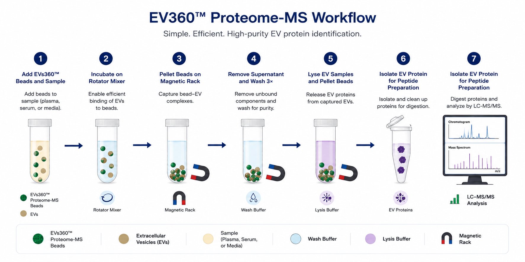
Protocol overview
Workflow
EVs360 Proteome-MS Kit protocol schematic
Product highlights
Instrument-free workflow
No ultracentrifuge needed — rapid isolation directly from plasma or serum
No secondary concentration
High yield without additional concentration steps required
In-depth proteomics
Optimized for high-depth blood EV proteomics and LC/MS workflows
Reproducible
Consistent performance across runs for quantitative proteomic studies
Automation ready
Compatible with HulaMixer and high-throughput liquid handling platforms
Order options
10 reactions
EVs360™ Proteome-MS Kit
Cat. no. L20001 · 10 rxn
List price$550.00
Per reaction0.25 mL plasma/serum
For useResearch use only
Protocols & manuals
Compatible downstream workflows
LC/MS proteomics workflows
High-sensitivity immunoassays (Olink, NuLISA)
Frequently Asked Questions

Practical guidance for EV enrichment and downstream protein biomarker and LC-MS/MS workflows.

What downstream applications is the kit designed to support?
The kit is designed for EV-associated protein workflows, including discovery LC-MS/MS, targeted mass spectrometry, Western blotting, ELISA, Olink®, NuLISA™, multiplex immunoassays, and protein biomarker validation. Confirm that the selected lysis buffer and cleanup method are compatible with the downstream platform.
What makes the workflow different from ultracentrifugation?
The kit uses lipid nanoprobe based magnetic beads and a 15-minute capture step, eliminating the need for an ultracentrifuge. Magnetic processing supports standardized handling, parallel sample preparation, and straightforward transfer to automation after workflow validation.
Which EV populations are captured?
The workflow provides broad, pan-EV enrichment rather than selecting a single EV subtype. The recovered material may include small EVs commonly described as exosomes, microvesicles, and other membrane-bound EVs. EV composition depends on sample type and pre-analytical handling.
What sample volume should I use?
The validated starting inputs are:
  • Plasma or serum: 250 μL per reaction.
  • Cell culture media: 2.5 mL per reaction.
The protocol may be scaled, but larger sample volumes are not a direct substitution. Bead amount, tube capacity, Solution A, wash volume, mixing, and magnetic separation must be optimized together. A larger input may increase total protein recovery, but proteome depth depends on EV abundance, background carryover, cleanup, digestion, instrument method, and data analysis.
Can frozen samples be used?
Yes. Frozen plasma, serum, and conditioned media can be used. Thaw completely, mix gently until homogeneous, avoid repeated freeze-thaw cycles, and use the pre-clear option in the Operator Manual if debris or aggregates are present.
What is the validated reaction setup for this kit?
Use 0.5 mL Beads L2 (2.5 mg) per reaction. Add 0.5 mL Solution A for plasma/serum; skip Solution A for cell culture media.
Does the kit require specialized equipment?
No ultracentrifuge is required. The workflow uses a magnetic stand, tube rotator or mixer, vortex mixer, calibrated pipettes, and a standard microcentrifuge. Use Protein LoBind tubes from capture through lysis to help minimize nonspecific protein loss.
What makes this kit suitable for blood proteomics?
Plasma and serum contain high-abundance proteins — including albumin, immunoglobulins, and lipoprotein-associated proteins — that can dominate proteomic measurements. The kit enriches an EV-associated fraction, and the three-wash workflow is designed to reduce soluble-protein and buffer carryover. This can support:
  • Improved access to lower-abundance EV-associated biomarkers
  • Greater proteome depth than direct analysis of unfractionated plasma or serum, depending on workflow and instrument performance
  • More consistent sample preparation across research cohorts
Is this an albumin- or IgG-depletion product?
No. The kit is not an albumin- or IgG-depletion product. Residual plasma proteins may remain, and results depend strongly on sample quality, wash consistency, lysis, cleanup, digestion, chromatography, mass spectrometry, and data analysis.
Which lysis buffers are recommended for LC-MS/MS?
Two starting options are described in the Operator Manual: 5% SDS with 10 mM TEAB or 8M Urea lysis buffer. Detergent-containing lysates require an MS-compatible cleanup and digestion workflow. After lysis, keep magnetic beads and residual Wash Buffer out of the transferred protein lysate.
Can this kit be used for biomarker discovery studies?
Yes. The workflow is designed to support discovery proteomics, translational biomarker studies, longitudinal research cohorts, and multi-center studies. For reproducible results, finalize the pre-analytical, capture, wash, lysis, cleanup, digestion, LC-MS/MS, and data-processing procedures before processing the full cohort.
Is the kit compatible with post-translational modification (PTM) proteomics?
Yes. EV protein lysates may be used in downstream PTM-enrichment workflows, including:
  • Phosphoproteomics
  • Glycoproteomics and glycopeptide analysis
  • Other PTM workflows after application-specific validation
PTM recovery depends on input, lysis chemistry, inhibitors, cleanup, digestion, enrichment, and MS sensitivity. Add appropriate phosphatase, protease, or other inhibitors for the selected workflow.
Is the workflow compatible with automation?
Yes. Magnetic separation, transfer, mixing, and washing are compatible with many liquid handlers. Validate bead resuspension, magnet time, aspiration height, format, mixing, carryover, and recovery.
What should I check if protein yield or proteome depth is lower than expected?
Review the following before changing the protocol:
  • Confirm sample input, thawing, mixing, and pre-clear conditions; vortex Beads L2 thoroughly.
  • Maintain continuous mixing during the 15-minute capture; use Solution A only for plasma/serum.
  • Complete all three 400 μL washes and remove the final wash completely.
  • Review lysis, inhibitors, cleanup recovery, digestion, LC-MS/MS performance, expected EV abundance, and biological variability.
Can captured material or protein lysate be stored?
For best consistency, proceed directly. If storage is required, transfer cleared lysate away from the beads and freeze at −80°C. Avoid repeated freeze-thaw cycles, document duration, and validate the pause point for the intended assay.
Is the kit suitable for clinical research studies?
The workflow can support translational, longitudinal, biobank, and multi-site research when the complete procedure is validated and standardized. The EVs360™ Proteome-MS Kit is For Research Use Only and is not intended for diagnostic procedures.
News and Recent Events

Latest from Captis
July 2026
Webinar Presentation with the EDRN
Captis Diagnostics presented "A Rapid Lipid Nanoprobe Platform for Extracellular Vesicle Isolation and Multi-Omics Diagnostics from Noninvasive Bodily Fluid" at a webinar in collaboration with the Early Detection Research Network (EDRN), a National Cancer Institute program dedicated to discovering, developing, and validating biomarkers for cancer risk assessment and early detection.
Visit EDRN →
March 2025
Captis Diagnostics Awarded NIH SBIR Phase II Grant to Advance Extracellular Vesicle Isolation Technology
Captis Diagnostics has received a Small Business Innovation Research (SBIR) Phase II grant from the NIH's National Center for Advancing Translational Sciences (NCATS) to advance its lipid nanoprobe (LNP) technology for rapid, highly efficient, and cost-effective EV isolation from non-invasive bodily fluids. Four research-use-only (RUO) EV isolation kits are now being prepared for commercial launch: Lipid Nanoprobe EV Isolation Kits for Plasma, Serum, Urine, and Saliva. The LNP platform supports comprehensive multi-omics characterization — genomics, transcriptomics, and proteomics — advancing biomarker discovery, precision medicine, and clinical diagnostics for a wide range of human diseases.
September 2023
Captis Diagnostics and Collaborators Receive Department of Defense Funding for Breast Cancer Research
Captis Diagnostics is pleased to announce that "Predicting Pathological Complete Response in Breast Cancer Using an Extracellular Vesicle-Based Liquid Biopsy" has been recommended for funding by the U.S. Department of Defense's Congressionally Directed Medical Research Programs (CDMRP) Breakthrough Award — Funding Level 1. In collaboration with researchers at Carnegie Mellon University, UPMC, and Penn State University, the project will investigate an EV-based liquid biopsy approach for predicting pathological complete response in breast cancer patients.
Company History
From Penn State Lab to Life Science Startup
Captis Diagnostics originated from extracellular vesicle (EV) research done at Penn State University, evolving from an academic research program into a Pittsburgh-based company. Today, Captis develops non-invasive, EV-based liquid biopsy tools for early disease detection and precision treatment guidance.

Meet Our Team

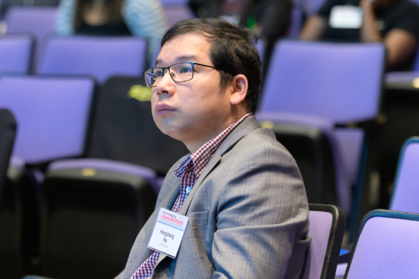
Hongzhang He
Hongzhang He
PhD · CEO & Co-Founder

Dr. He is a medical diagnostics entrepreneur with significant expertise in EV isolation and molecular diagnostic assay development. He has received extensive training in business development through TechCelerator, NIH I-Corps, LifeX Lab, and NSF I-Corps. He has focused on developing EV-based minimally invasive technology for human disease diagnosis, along with ddPCR and next-generation sequencing molecular diagnostic assays. Dr. He has authored over 50 publications with 3,675 citations on Google Scholar, and holds 1 issued patent with 5 patent applications.

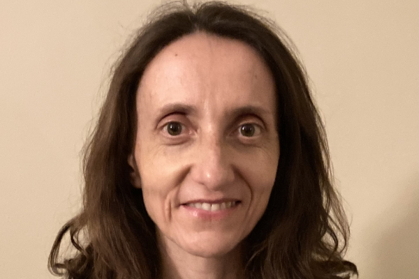
Jadranka Milosevic
Jadranka Milosevic
PhD · Principal Research Scientist

Dr. Milosevic is an accomplished researcher and R&D scientist with extensive experience in epigenetics, biomarker discovery, and EV research. Her primary objective is to develop minimally invasive liquid biopsy tools as routine diagnostic tests for human disease. Over more than 15 years, she has acquired substantial expertise and has authored 25 scientific publications. She has played various roles from mentoring junior scientists to leading independent and collaborative research.

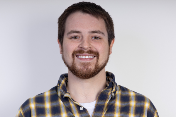
Brandon Park
Brandon Park
PhD · Research Scientist I

Dr. Park is a molecular biologist and bioinformatician with expertise in next-generation sequencing, epigenomics, and extracellular vesicle research. He completed his Ph.D. in Genetics at the University of Rochester School of Medicine and Dentistry, where his doctoral work focused on developing novel genomic technologies including Cas-CUT&Tag and CUT&Tag-based epigenomic profiling. At Captis Diagnostics, he leads development of LEVR-Seq (long-read Nanopore sequencing for EV-derived RNA isoform and gene fusion detection) and EV-EPIC (CUT&Tag epigenomic profiling from EV-derived chromatin), and serves as Principal Investigator on NIH SBIR-funded projects.

We're building
something meaningful

Captis Diagnostics is a growing team working at the intersection of nanotechnology, oncology, and molecular diagnostics. If you're passionate about making minimally invasive disease detection a clinical reality, we'd love to hear from you.

Get in Touch

How can we help?

Further information available upon request. Please provide some details on your project or goals and we'll move the conversation forward from there.

Address
4620 Henry Street
Pittsburgh, PA 15213
Phone
+1-949-878-2679
Hours
Mon–Fri, 9:00–17:00 ET
Sat–Sun closed